SHENZHEN besar PENCIPTAAN TEKNOLOGI TERBATAS

Apakah kita yang terbaik bagi pelanggan kami lebih baik!

Rumah
Produk
Tentang kita
Wisata pabrik
Kontrol kualitas
Hubungi kami
Quote request suatu
Rumah ProdukMesin Ultrasound Ponsel

4D Color Doppler Ultrasound Scanner with 3.5MHz / R40 Volume Probe

kualitas baik Quantum magnetic resonance kesehatan analyzer untuk penjualan
Excellent Shipping. very fast response to questions AAA+++++ seller will do business with again.

—— Jim.M

Super venditore,affidabile,veloce sulla spedizione.....insomma tutto ok:-)

—— Giovanni S.

Thank you. I am very happy with the product. Everything arrived in excellent condition.

—— Rayner L

quality Quantum Magnetic Resonance Health Analyzer. Ok trade.

—— Ricardo Jorge

todo bien excelente producto y excelente vendedor siempre muy cordial y en comunicacion

—— Lopes Milena

I 'm Online Chat Now

4D Color Doppler Ultrasound Scanner with 3.5MHz / R40 Volume Probe

Cina 4D Color Doppler Ultrasound Scanner with 3.5MHz / R40 Volume Probe pemasok
4D Color Doppler Ultrasound Scanner with 3.5MHz / R40 Volume Probe pemasok 4D Color Doppler Ultrasound Scanner with 3.5MHz / R40 Volume Probe pemasok 4D Color Doppler Ultrasound Scanner with 3.5MHz / R40 Volume Probe pemasok 4D Color Doppler Ultrasound Scanner with 3.5MHz / R40 Volume Probe pemasok

Gambar besar :  4D Color Doppler Ultrasound Scanner with 3.5MHz / R40 Volume Probe

Detail produk:

Place of Origin: China
Nama merek: Huge
Sertifikasi: CE
Model Number: EC-50D

Syarat-syarat pembayaran & pengiriman:
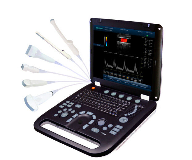
Minimum Order Quantity: 1
Harga: USD$7500/pcs
Packaging Details: carton box
Delivery Time: 3-5 working days after get payment
Payment Terms: MoneyGram, Western Union, T/T,paypal
Detil Deskripsi produk
product name: 4D Color Doppler Ultrasound Scanner Carton size: 47*48*29 cm
Net weight: 5 kgs Gross weight: 8 kgs
standard probe: 3.5MHz / R40 Volume Probe
Cahaya Tinggi:

personal ultrasound machine

,

equine ultrasound machine

4D Color Doppler Ultrasound Scanner with 3.5MHz / R40 Volume Probe


Quick Detail:


Color Doppler (CFM)

Power Doppler (PDI)

Directional Power Doppler (DPDI)

Pulsed Wave Doppler (PWD)

continuous wave doppler CW

B+PWD (Duplex)

B+CFM/PDI/DPDI+PWD (Triplex)

High Pulse Repetition Frequency (HPRF)

Tissue Harmonic Imaging (THI)


Description:


Ultrasound diagnosis technique is to use the ultrasonic as information carrier, the ultrasonic probe transmits ultrasonic to human body, and through the same probe (or called as transducer)receive echo with information on human body tissue, then through information extraction and processing to realize the inspection and diagnosis on human body tissue, it has some advantages in safety, no wound, direct viewing, real time, repeatable inspection, convenient operation, wide application, inexpensive price and stronger discriminability to parenchyma. It has taken possession of very important position at the current four image diagnosis techniques in medicine, it has been widely applied to clinic diagnosis, family planning and getting well and health protection, the ultrasonic diagnostic instrument has become a popular and conventional diagnosis system.

4D Color Doppler Ultrasound Scanner with 3.5MHz / R40 Volume Probe


Applications:

  • Abdomen section (adults and pediatrics) : to detect abnormalities from the structure images of liver, kidney, pancreatic gland, gall bladder, spleen, gastrointestinal tract and urogenital organ.
  • Abdomen section (obstetrics and gynecology): to detect and discover abnormalities from the structure images of fetus, womb and pelvis so as to estimate the fetal age and weight and evaluate the fetal heart function.
  • Chest section (adult and pediatric): to analyze M-mode image so as to detect abnormalities of the heart structure and function.
  • Skin section (organella):to detect abnormalities from the rough evaluation of images of breasts, thyroid, and testicles, etc.
  • Skin section (peripheral vessel): to detect and evaluate the vascular stenosis and occlusion from the images of peripheral vessels and flow measurement.
  • Neonatal head section: to detect abnormalities of the cerebral volume and structure from the image of neonatal head.
  • Urological section: to detect the left/right kidney, bladder and calculate residual urine volume, etc.
  • Musculoskeletal section: to detect and discover abnormalities from the structure images of fetus and musculoskeletal in the application of obstetric and gynecology.
  • Transvaginal section: to detect and discover abnormalities from the structure images of uterus, ovary, ovarian follicular, etc.


Specification:


Standard Configuration


1. EC-50D main unit

2. 3.5MHz/R40 volume probe

3. 4D imaging software package

4. phased array probe

5. 250ml coupling gel

6. AC adapter

7. Built-in rechargeable lithium battery

8. Protection bag

9. Grounding cable

10.Fuse (2)

11. Operation manual


Image:

B, B|B, 4B, B|M, M,4D
Color Doppler (CFM)
Power Doppler (PDI)
Directional Power Doppler (DPDI)
Pulsed Wave Doppler (PWD)
continuous wave doppler CW
B+PWD (Duplex)
B+CFM/PDI/DPDI+PWD (Triplex)
High Pulse Repetition Frequency (HPRF)
Tissue Harmonic Imaging (THI)

4D Color Doppler Ultrasound Scanner with 3.5MHz / R40 Volume Probe

4D Color Doppler Ultrasound Scanner with 3.5MHz / R40 Volume Probe


Rincian kontak
Shenzhen Huge Creation Technology Limited

Kontak Person: Ms. Karen

Mengirimkan permintaan Anda secara langsung kepada kami (0 / 3000)